
What A CBCT Scan Shows In One 3D Image
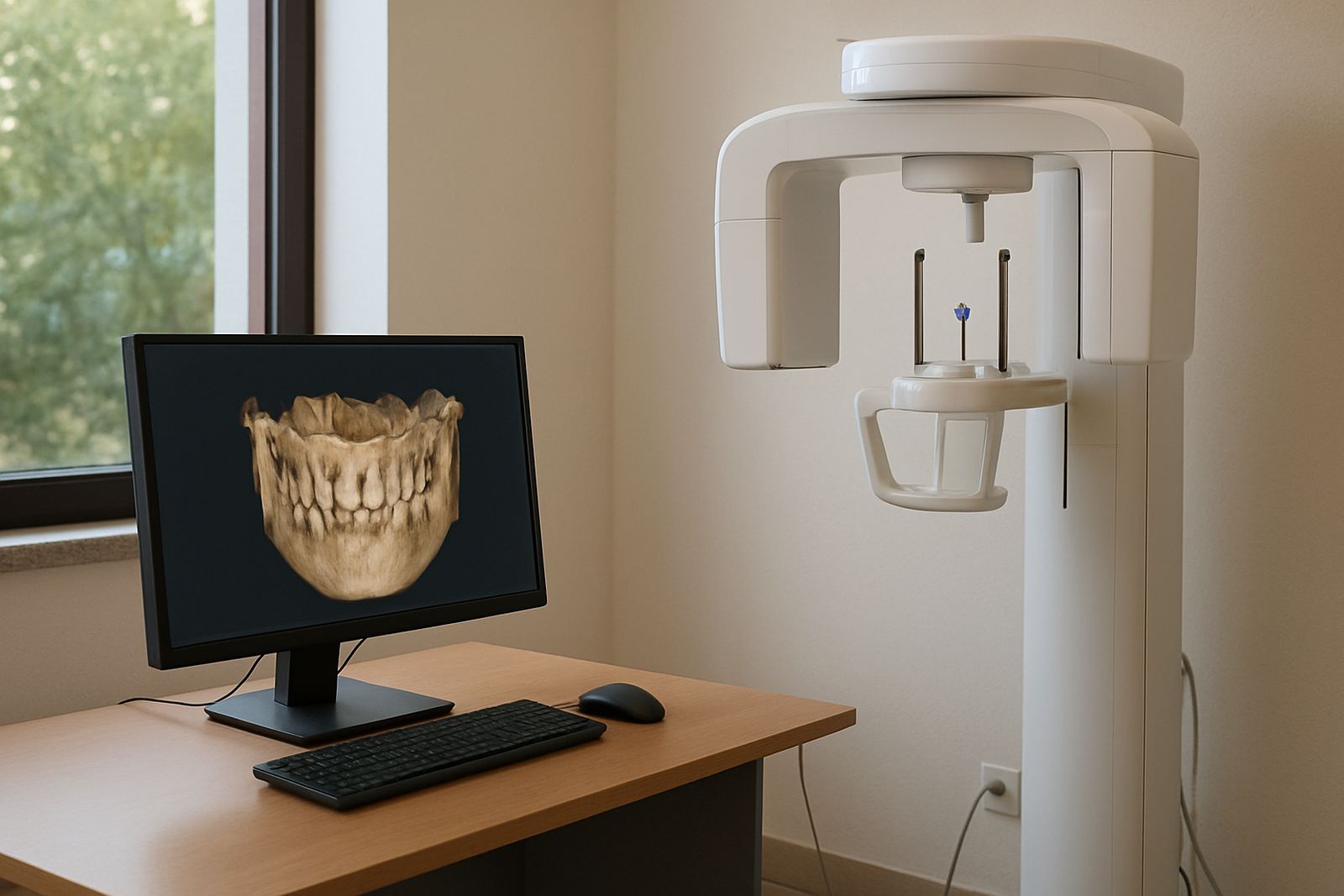
A CBCT scan, or cone beam computed tomography, gives a three-dimensional view of the teeth, jaws, airways, nerve paths and surrounding bone. It is used when a normal dental X-ray does not show enough detail for treatment planning, especially where exact depth, angle and bone volume matter.
For implant cases, the scan helps the clinician measure bone height and width, locate the sinus floor or nerve canal, and plan the implant position before any procedure starts. For more complex cases, it can show impacted teeth, root anatomy, infection spread, cysts, fractures and joint-related concerns much more clearly than a flat image.
At Techno-Dent Clinic, CBCT imaging is reviewed alongside the clinical examination, photographs and treatment goals so the scan is not just a picture, but part of a proper plan.
When CBCT Imaging Is The Right Choice
CBCT is usually requested when precision matters and the case needs more than routine bitewing or panoramic imaging. It is especially useful before implants, for orthodontic assessments, for wisdom teeth close to nerves, and for evaluating complicated root canal problems.
- Implant planning where bone dimensions must be checked before surgery
- Orthodontic assessments for impacted teeth or jaw relationships
- Emergency dentistry when trauma or fractures need clearer imaging
- Cosmetic dentistry cases where alignment or support structures need review
- Aftercare follow-ups when healing or bone changes must be monitored
In Sandton and the surrounding areas, many patients come in after a referral or after their own dentist has asked for a more detailed scan. That helps avoid guesswork and can save time later in treatment.
Scan Size, Field Of View And Detail Levels
CBCT systems are not one-size-fits-all. The field of view, or scan area, can be selected according to the case. Smaller views are used for a few teeth or a single implant site, while larger views are used for both jaws, the full mouth or airway-related assessments.
Common scan options include a small field of view for endodontic or localised planning, a medium field for single arch studies, and a large field for full jaw or facial analysis. The correct setting keeps radiation exposure as low as practical while still giving enough detail for the clinical decision.
- Small field of view: localised detail for one or a few teeth
- Medium field of view: partial arch planning and local surgical checks
- Large field of view: full jaw, both arches or airway-related review
- High-resolution mode: finer detail for roots, canals and bone edges
For many patients, the scan takes only a short time and the images are available immediately for treatment discussion. If you are nervous in the chair, the team can explain the process step by step before starting.
How CBCT Supports Implant Planning And Surgery
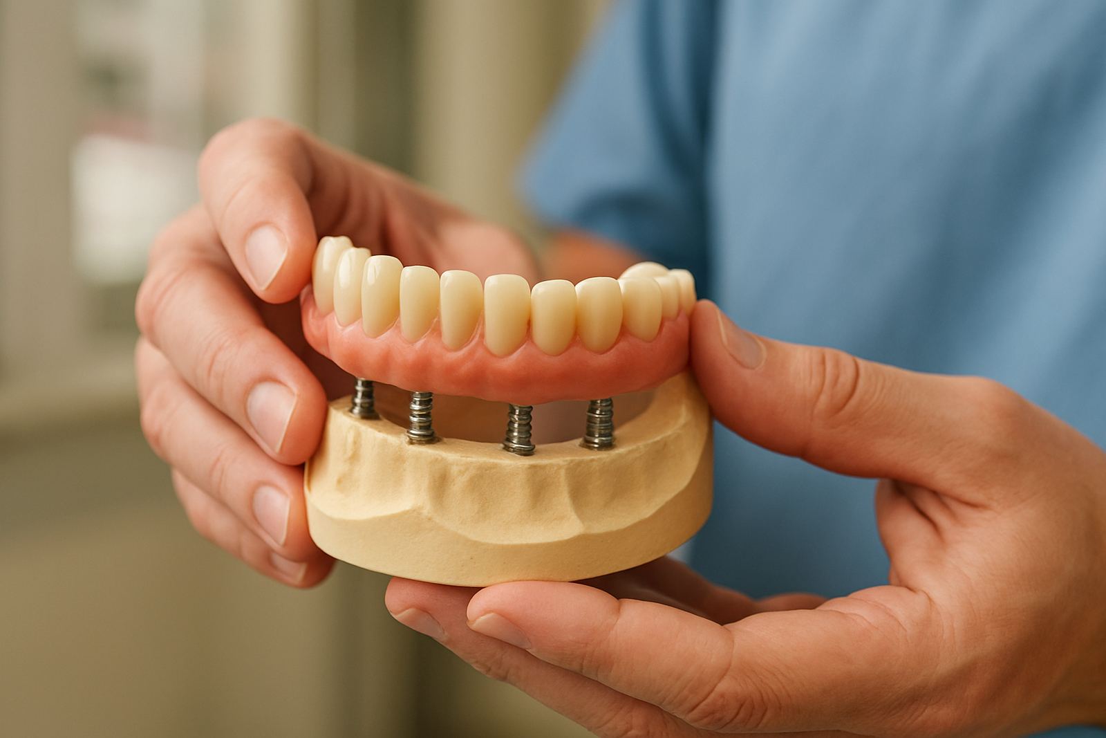
Implant dentistry depends on measurements. A CBCT scan gives the clinician the data needed to choose implant length, width and placement angle with far more confidence. It also helps identify where extra procedures may be needed, such as bone grafting or sinus lift planning, before the day of surgery.
For surgical work, the scan improves visibility of important structures like nerves, roots and sinus spaces. That means a more careful plan and better control during treatment. In orthodontics, the same 3D detail can help identify the exact position of unerupted teeth and guide whether referral, exposure or monitoring is best.
Dr Pieter van der Merwe and Dr Nandi Dlamini work with detailed imaging findings to support treatment choices for implants and orthodontic cases. This is especially useful when the anatomy is unusual or the case needs proper pre-treatment planning rather than a quick look.
Installation Needs, Power And Practical Site Requirements
CBCT equipment needs a dedicated room with controlled access, suitable wall space and stable flooring. The room should allow for patient positioning, safe movement around the unit and enough space for staff to operate without crowding. Good lighting and clear signage help patients move through the appointment without confusion.
Power requirements are typically standard mains electricity with a stable supply, and a UPS or backup power solution is wise in Johannesburg where load-shedding can interrupt clinical flow. Surge protection is important too, because imaging equipment is sensitive to voltage spikes. Some practices also plan for data backup and network support so scans can be stored and retrieved reliably.
- Dedicated room with enough clearance around the unit
- Stable mains power and surge protection
- Backup power for outages and load-shedding periods
- Controlled access for patient privacy and safety
- Workflow that supports image storage and review
There are no gas requirements for CBCT imaging. What matters is a tidy clinical layout, proper electrical support and enough ventilation for patient comfort, especially in warm inland months or in rooms that run for most of the day.
Care, Servicing And Image Quality Over Time
CBCT systems need routine servicing to keep image quality consistent and to make sure positioning, calibration and software functions stay accurate. Dust control matters, as do clean screens, correct positioning aids and regular checks on the machine components used for scanning.
The team should also keep an eye on infection control, patient flow and record keeping. If the scan room is used often, daily wipe-downs and weekly checks help prevent small issues from turning into downtime. In coastal humidity or dusty periods, simple maintenance discipline goes a long way.
At Techno-Dent Clinic, CBCT imaging is supported by a digital workflow coordinated by Thabo Nkosi, so scans can be managed, reviewed and shared with the treating clinician without unnecessary delay.
What To Check Before You Order Or Book A Scan
Before booking a CBCT scan, it helps to confirm exactly what the clinician wants to see and how large the scan area should be. A scan that is too small may miss important anatomy, while one that is too large may include unnecessary detail and extra exposure.
- Requested field of view and whether one jaw or both jaws are needed
- Whether high-resolution images are required for canals, roots or implants
- Patient positioning needs, including mobility or sedation considerations
- Delivery format for the images and report
- Clinical purpose, such as implant planning, trauma review or orthodontic assessment
- Timing for the scan and any follow-up appointment needed
If you are a patient in Sandton, Bryanston, Rivonia, Morningside, Fourways or Rosebank, the practice can guide you on the scan process and payment questions before the appointment. For urgent cases, emergency dentistry and new patient consults can be arranged with the imaging request included where needed.
Key facts
| Scan type | Cone beam computed tomography, 3D dental imaging |
|---|---|
| Typical field of view | Small, medium and large options for localised to full jaw studies |
| Main uses | Implant planning, complex diagnosis, surgical precision, orthodontic assessment |
| Image output | 3D DICOM imaging with cross-sectional views and measurement data |
| Power need | Stable mains electricity with surge protection and backup power recommended |
| Gas need | None required |